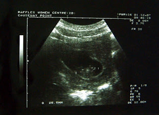
Today we went for the DS (Detailed Scan) at Raffles Hospital and several measurements were made to detect structural anomalies. In the pic above the baby's Biparietal diameter was measured (Skull).
Everything was great and we found out that it was a baby GIRL.
As of current the EDD is 30 June 2008
What is examined at the 20 week scan?
The baby's internal organs are examined in cross-sectional views.
Bones appear white on the scan, fluid is black, and soft tissues look grey and speckled.
The head is usually examined first, to check its shape and internal structures.
Some severe brain problems are visible at this stage. The sonographer will look at the baby’s face to see if she can see any cleft of the lip, but clefts of the palate inside of the baby’s mouth are difficult to see and are not often picked up on scans.
The spine is checked in both the long view and in cross section, to make sure that all the vertebrae are in alignment and that the skin covers the spine at the back.
The baby's abdominal wall is also checked to make sure it covers all the internal organs at the front.
The heart is looked at for its size and shape. The top two chambers, or "atria", and the bottom two chambers, or "ventricles", should be equal in size, and the valves should open and close with each heartbeat.
Raffles offer a more detailed scan of the heart to look at the major blood vessels.
The stomach should be visible below the heart.
The baby swallows some of the amniotic fluid that it lies in, which is seen in the stomach as a black bubble.
The sonographer will check that the baby has two kidneys, and that urine flows freely into the bladder. If the bladder is empty it should fill up during the scan and be easy to see - the baby has been weeing every half an hour or so for some months now!
Hands and feet are examined and the fingers and toes are looked at, but not counted.
The placenta, umbilical cord and amniotic fluid are all checked The placenta may be on the front or the back wall of your womb, usually near the top (or fundus) so may be described as "fundal" on your scan report. Many are described as "low" because they reach down to or cover the neck of the womb (cervix).
If your placenta is low, another scan will be arranged in the third trimester, by which time most placenta will have moved away from the cervix.
It is possible to count the three blood vessels in the umbilical cord, but this may not be done routinely. There should be enough amniotic fluid surrounding the baby to allow it to move freely at this stage.
Next update Mid March 2008~


